Sunday, 17 January 2016

GASTRO INTESTINAL FOREIGN BODIES IN DOGS


The gastrointestinal tract consists primarily of the stomach, small intestine and large intestine. It is always difficult to see dogs suffering from foreign body ingestion. But as a Surgeon much excitements and rewarding results are seen in immediate intervention of foreign body ingestion cases . The foreign body entered in side the mouth can pass smoothly through the oesophagus , stomach , small intestines- duodenum, ileum, jejunum, Large intestines- cecum colon rectum and finally it can come through the anal opening outside . But it can also get struck up in any place and the commonest blocks can be encountered in
1.      Pharynx – Isthmus faucum
2.      Oesophagus
3.      Stomach
4.      Duodenum
5.      Jejeunum
6.      Ileum
7.      Ileo cecal junction
In this blog I am sharing some memorable cases of intestinal obstruction in my career.
Due  to indiscriminate eating habits from PICA or Phosphorous deficiency, high worm load due to improper deworming or accidental ingestion of toys or plastic or hard objects , the animal may swallow hard foreign objects which can lodge at any of the above mentioned sites 


NON LINEAR OR SOLID  FOREIGN BODY 
Solid foreign bodies may get lodged in any part of the GIT depends up on the sharpness of edges , size and consistency. There were very two very interesting cases of non linear foreign bodies last month.
TOY WHEEL FOREIGN BODY IN THE JEJUNUM OF A  LABRADOR RETRIEVER 

 A Labrador was referred by a field veterinarian in Thrissur with the history of off feed, not passing motion and extremely painful abdomen. On palpation a hard mass was palpable on the cranial abdomen caudal to the stomach .close examination of  lateral abdomen radiograph revealed a  shadow of  round mass won the abdomen caudal to stomach . On exploratory laparotomy a toy wheel was retrieved from jejunum.  


GRANITE STONE IMPACTION IN A BASSET HOUND

A six year old male Basset hound was presented with symptoms suggestive of chronic pica from the past one month of presentation . The animal was cachectic , anorectic  and was showing recurrent vomiting signs non responsive to medical treatment. On examination a hard mass was palpable on the ventral abdomen and plain radiography revealed a radio opaque mass on the cranial abdomen obliterating the entire lumen and gas filled loops caudal to the obstruction. The animal was stabilised with supplementary fluid therapy
. An exploratory laparotomy was performed by induction and maintenance using thiopentone sodium 2.5 % to the effect under general anaesthesia with atropine and Xylazine as pre medicants @ 0.045 mg /kg bw and and 1 mg / kg bwt respectively.  A hard intestinal segment was isolated at the mid jejunum and a 2 cm long enterotomy incision on non-mesenteric border exteriorised a granite stone piece was exteriorised. Enterotomy closure was carried out using  No 2-0 polyglactic acid suture material in close interrupted pattern. Linea alba was apposed using No 1 poly glactic acid 910 in simple interrupted pattern followed by skin closure in routine manner . Post operatively the animal was treated with antibiotic therapy and fluid therapy for 5 days and oral feeding was withheld for 72 hours post surgery. Sutures were removed on 11th post-operative day and the animal had uneventful recovery.


LINEAR FOREIGNBODY
Gastrointestinal foreign bodies are regarded as main GI emergencies in companion animal practice .Linear foreign bodies, especially strings, can often lead to extreme painful condition and can result in intussusceptions and volvulus which demands immediate surgical intervention.

STORY OF DISCO
I had a dog named Disco when I was working at Hassan Veterinary College. Disco use to accompany me where ever I go and once he came running to me with a long thread seen protruding from his anal opening. On examination a large dressing gauze that he swallowed from the clinics was detected coming out of anus.. One of the student with a good intention to relieve his struggle pulled the thread and he was  crying out of pain .
An immediate laparotomy was performed and a large knot of gauze leading to intussusception (invagination of one part of intestine – intusussipiens to the other part – intususceptum was identified) . The intussusception was corrected and we saved Disco by a timely surgery  

YOU CAN WATCH A VIDEO ON GASTRO INTESTINAL FOREIGN BODIES BELOW 


SIGNS OF OBSTRUCTION
1.    Ingested foreign bodies are more common in young animals                                        
2.    Vomiting is usually seen with an obstructive foreign body of the gastrointestinal tract, however this is a problem that may be seen with many other diseases                                     
3.    Generally if an obstruction of the GI tract is present, diarrhea is usually not a common symptom (as the intestines are obstructed)                                                                             
a.       Loss of appetite                                                                                                                      
b.      Dehydration                                                                                                                               
c.       Abdominal pain                                                                                                               Weight loss may be present if the foreign body is chronic
DIAGNOSIS
1.    Haematology  work is done to determine if there are liver, kidney, pancreas, or electrolyte abnormalities. Complete blood cell count is used to look for signs of infection and anemia.
2.    Radiographs (x-rays) can be very helpful to indicate the presence of a problem in the gastrointestinal tract. Unfortunately, plain radiographs frequently are only suggestive of a problem and do not give us a definitive answer; in some cases the radiographs will be made again to see if the signs of obstruction are repeatable. As a result additional tests such as ultrasound or contrast radiographs (barium swallow) may be indicated.
3.    Sometimes these tests also do not give us a final answer and exploratory surgery is needed.              
4.    If we do not find any obvious problem with the internal organs, biopsies still are done as microscopic disease may be causing the clinical signs.

PRINCIPLES OF INTESTINAL SURGERY
1. Early diagnosis and good surgical technique prevent most complications.
2. Perform surgery as soon as anesthesia is possible in patients with perforation, strangulation, or complete obstruction.
3. Optimal healing requires a good blood supply, accurate mucosal apposition, and minimal surgical trauma.
4. Systemic factors may delay healing and increase the risk of dehiscence, hypovolemia, shock, hypoproteinemia, debilitation, and infection.
5. Use approximating suture patterns: simple interrupted, Gambee, crushing, or simple continuous; stapling techniques are feasible.
6. Engage submucosa in all sutures or staples.
7. Select a monofilament, synthetic absorbable suture such as polydioxanone, polyglyconate,
8. Cover surgical sites with omentum or a serosal patch.
9. Replace contaminated instruments and gloves before closing the abdomen.
10. Fluid Therapy
11. Antibiotic Prophylaxis
12. Assessment of Intestinal Viability
13. Choice of suture material for closure.
14. Monofilament synthetic absorbable (PDS) or synthetic non-absorbable (prolene) are excellent choices.
15. Choice of Suture Pattern
16. Simple interrupted pattern is ideal.
17. Suture Reinforcement
18. Application of Omental and serosal patch aids in faster healing
19. Obtain minimum database: complete blood cell count, chemistry profile, urinalysis, coagulation profile (if possible) with or without an electrocardiogram.
20. Localize the lesion with abdominal palpation radiographs, ultrasonography, and/or endoscopy. Correct hydration, electrolyte, and acid-base abnormalities. Transfuse if the packed cell volume is less than 20% or if the animal is clinically weak or debilitated .Withhold food from mature animals for 12 to 18 hours and from pediatric patients 4 to 8 hours before induction.


 THANKS FOR VISITING MY BLOG 

Friday, 23 October 2015

CANINE MAMMARY TUMOR

This segment is intended to give an insight on canine mammary tumors its  direct correlation of the intact uterus and ovaries and the  effect of early  neutering in dogs .

In our university hospital on an average we receive 5-8 cases per week presented with advance canine mammary tumors  . It is a great concern for the dog owners as there is a large awareness among the cancer in human beings. But it is a sad reality that even the most educated pet owner is not fully aware of the direct correlation between the mammary tumor and the intact uterus and ovary in dogs . 

CANINE MAMMARY GLAND - ESSENTIAL  ANATOMY
The number of teats in the dog varies from 8 to 12, with 4 to 6 gland complexes on each side of the midline. Ten is the most common number in larger breeds, four pairs are more common in the smaller breeds. In bitches with ten normal teats, the pattern is two pairs thoracic teats, two pairs abdominal teats, and one pair of inguinal teats.


The number of ducts opening on a teat varies from 8 to 20 external openings per teat for the dog and 1 to 7 for the cat. The openings are located on the blunt end of the teat in an irregular pattern.

The parenchyma, or secretory tissue, is present only during pregnancy, pseudopregnancy, during lactation, and for 40 to 50 days after weaning.
The blood supply of the mammary glands of dogs and cats are similar except for the thoracic glands. In the dog the first pair of thoracic mammary glands receives blood from two sternal branches of the internal thoracic artery, passing between the first and second ribs. The second pair of thoracic mammary glands is supplied by small branches of the mediastinal, or internal mammary, arteries before they anastamose with the sternal branches serving the first pair of glands.

MAMMARY TUMORS
Tumors are frequently seen in the mammary gland of the dog. They may belong to the connective tissue or the epithelial series of mammary tumors, or both. Tumors of the epithelial series are of great importance. Those observed are adenomas, carcinomas, and above all, mixed mammary tumors.
The risk of breast cancer is almost eliminated in dogs that are spayed before their first heat.
Spaying greatly reduces the chances of a female dog developing this condition.
 In those females spayed prior to their first heat cycle, breast cancer is very, very rare. The risk of malignant mammary tumors in dogs spayed prior to their first heat is 0.05%. It is 8% for dog spayed after one heat, and 26% in dogs spayed after their second heat. It is believed that the elimination or reduction of certain hormonal factors causes the lowering of incidence of the disease in dogs that have been spayed. These factors would probably be estrogen, progesterone, a similar hormone or possibly a combination of two or more of these.

The development of mammary gland neoplasms appears to be hormone-dependent because the risk of developing a mammary tumor increases as the number of estrous (heat) cycles increases.

Normal mammary tissue and a majority of benign tumors express both estrogen and progesterone receptors. Less than 50% of mammary carcinomas express either of these receptors. This observation suggests that there is a loss of hormone dependency during transition to malignancy.

Common Signs
Abnormal swellings that persist or continue to grow
Sores that do not heal
Weight loss
Loss of appetite
Bleeding or discharge from any body opening
Offensive odour
Difficulty eating or swallowing
Hesitation to exercise or loss of stamina
Persistent lameness or stiffness
Difficulty breathing, urinating, or defecating

Mammary carcinomas may exhibit rapid growth, doubling in size within a few weeks. However, the size and appearance of these neoplasms can vary greatly. Inflammatory carcinomas usually have diffuse involvement of multiple mammary glands. Edema, erythema, and firmness may be present and affected mammary glands may feel warm to the touch. Dogs with inflammatory carcinoma are more likely to have generalized weakness with anorexia and weight loss. Inflammatory carcinoma is often misdiagnosed as acute mastitis.

TYPE OF TUMORS 
The following are common classes of mammary tumors that might be found on a biopsy.
Fibroadenoma:
A benign glandular tumor for which no treatment is necessary.
Mixed Mammary Tumor:
What is mixed is the type of cell that makes up the tumor: the epithelial cells that line the glandular tissue and the mesenchymal cells that make up the non-glandular portion. (Mixed does not refer to a mix of benign and malignant cells.) The mixed tumor can be either benign or malignant and the biopsy will indicate this.
Adenocarcinoma: 
Adenocarcinomas can be tubular or papillary, depending on the gland cells the tumor arises from. Adenocarcinomas behave malignantly but how aggressively malignant they are depends not on whether they are tubular or papillary, but on other cellular characteristics described by the pathologist (such as how quickly the cells appear to be dividing and how closely they resemble normal gland cells). When the oncologist reads the description he or she will be able to determine how aggressively to combat the tumor.
Inflammatory Carcinoma:
A highly malignant tumor that generates tremendous inflammation locally with ulceration, pus, and discomfort. This type of tumor tends to spread early in its course and is difficult to treat. Fortunately, this especially tragic tumor type accounts for less than 5% of mammary tumors.
In general: approximately 50% of malignant mammary tumors will have already spread by the time of surgery.
The malignant mammary tumors thus in general includes
Tubular adenocarcinomas
Papillary adenocarcinomas
Papillary cystic adenocarcinomas
Solid carcinomas
Anaplastic carcinomas
Osteosarcomas
Fibrosarcomas
Malignant mixed tumors.

Diagnosis of Mammary tumors
Mammary gland tumors are difficult to diagnose by routine cytology and the malignant potential of mammary neoplasms cannot be easily detected in early stages by cytologically.

The cytologic criteria for malignancy in Mammry tumors are

Anisocytosis (variable nuclear size)
Nuclear giant forms
Nuclear or cytoplasmic membrane distortions
High nuclear to cytoplasmic (N:C) ratio
Irregular chromatin shape
Variable chromatin size
Presence of macronucleoli
Variation in nucleolar number
Variation in nucleolar shape
Parachromtin clearing

Hormones 
Approximately 50% of malignant mammary tumors in the dog have receptors for either estrogen or progesterone. This means that the presence of these female hormones promotes the growth of these tumors. Benign tumors also have female hormone receptors and can also be stimulated by hormonal cycling of the female dog. This means that spaying is important even if a tumor has already developed.

MANAGEMENT OF CANINE MAMMARY TUMOR 
Surgical removal of the tumor(s) is the treatment of choice for benign mammary tumors and for malignant mammary tumors that have not spread beyond the mammary tissue and adjacent lymph nodes. If  surgery is done early in the course of this disease, the cancer can be totally eliminated in over 50% of the cases having a malignant form of cancer.Most benign mammary tumors are curable by surgery. Approximately half of malignant mammary tumors are also cured by surgery. This is possible because some malignant mammary tumors in the dog do not spread very quickly and can be removed before they spread. Radical mastectomy has not been shown to be any more effective than more limited surgery. Tumors that are larger than 3 centimeters and tumors that are of higher grade (as classified by the pathologist) are more likely to recur (70% recurrence at 1 year) than smaller tumors and tumors of a lower histopathologic grade (30% recurrence at 1 year). If the dog is not spayed at a young age, spaying at the time of mass removal has recently shown to increase the survival time for some dogs with malignant tumors.
Different terminologies are related to the surgical resection of mammary gland

Mammectomy
Removal of one entire mammary gland, indicated for lesions involving 1/3 of the gland or that are fixed to skin or fascia. Remove skin, abdominal wall fascia.

Regional mastectomy
Removal of affected gland and adjacent mammary glands based on known venous/lymphatic drainage of mammary tissue (may involve axillary, superficial inguinal, sublumbar and cranial sternal nodes).

En bloc mastectomy
Resection of affected glands with the regional lymph node(s)

Unilateral or bilateral (radical) mastectomy
Indicated if multiple tumors or several large tumors along one or both mammary chains, or for any malignant mammary gland tumor in cats.


Chemotherapy: An effective chemotherapy protocol for canine mammary cancer has not been defined. A small percentage of dogs have had remission with drugs such as doxorubicin (Adriamycin) or cisplatin. Because surgery alone is successful in many cases, chemotherapy is usually reserved for tumors that cannot be removed surgically, tumors that have metastasized, and tumors that have a high probability of spreading. Radiation therapy has not been extensively studied to in dogs with mammary tumors. The chemotherapeutic protocol which  can be tried will be a combination of 5-fluorouracil (150 mg/m2 of body surface  area) and cyclophosphamide (100  mg/m2)  but the sideffects masks the results. Postoperative Adjuvant Treatment of Invasive Malignant Mammary Gland Tumors in Dogs with Doxorubicin and Docetaxel has also given excellent results for many practitioners .
A word of caution is always advisable using doxyrubicin . In one case I had encountered a very adverse reaction and tachy cardia associated with doxyrubicin.
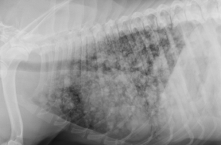
Metastasis is a common feature of malignant mammary tumors and chest radiographs will reveal the metastatic nodules in lungs in most of the advanced carcinomas. 
Some cases give real challenges with multiple lesions 
PLEASE WATCH THE VIDEO BELOW 


PREVENTING MAMMARY TUMORS


There is a direct correlation between the early spaying of female dogs and the reduction in the incidence in mammary cancer.
Dogs spayed before coming into their first heat have an extremely small chance of ever developing mammary cancer. 
Dogs spayed after their first heat but before 2.5 years are at more risk, but less risk than that of dogs who were never spayed, or spayed later in life. As a veterinarian I strongly believe that .......MAMMARY TUMORS ARE  PREVENTABLE  BY EARLY SPAYING . 

Saturday, 18 July 2015

FELINE DYSTOCIA AND CESAREAN SECTION


I am sharing some insights on the Feline Dystocia and C- Section in this segment .
Many times we get cats presented in advanced stage of dystocia. It is very important for the cat owners, Paravets to understand the stage of birth which clearly indicates the time for intervention by the vet.
Cat’s labour signs are very interesting and much more unique compared to canine labour stages .

STAGE -1 First stage labour begins with the onset of uterine contractions. During this stage the cervix begins to dilate open with clear , odourless discharge from the vagina - mucus plug during pregnancy sealing the uterus starts. As the first stage progresses, contractions will become closer and closer together.

STAGE -2 Second stage labour is characterised by contractions much stronger and closer together and the cervix becomes fully dilated. When the queen is ready , kitten moves down the birth canal. Pressure   on the cervix initiates an urge to push from the mother. It takes around three pushes for the kitten to be delivered. The queen will tear and lick the membranes from the face and body which will stimulate breathing. Second stage usually takes around 5 minutes to 1 hour.
MORE THAN ONE HOUR OF SECOND STAGE OF LABOUR NEED VETERINARY HELP!

STAGE -3 Third stage labour: Immediately following the kitten's birth, the placenta is normally delivered. Once the queen has cleaned the kitten and breathing has commenced normally the queen will chew the umbilical cord in two and quite often will eat the placenta.

CAUSES OF DYSTOCIA IN CAT
 There  are a number of causes of dystocia which can be classified as maternal or fetal. Maternal causes include  Narrow pelvis. Fractured pelvis ,Pelvic obstruction and Uterine inertia uterine torsion (twisting) or rupture. This may be primary or secondary . When the uterus fails to produce any contractions this is regarded as Primary inertia This can be from  poor health, obesity, age, stress.Fetal causes include abnormally presentation of  kitten, oversized foetus , Excessive fetal head size and malformed fetus, Fetal death.

A CASE OF UTERINE TORSION LEADING TO DYSTOCIA AND REVIVAL OF LIVE KITTEN BY CESAREAN SECTION 
PLEASE WATCH THE VIDEO BELOW .


We had handled a very challenging case of feline dystocia this week. three year old cat was presented on 71 day of gestation with history of delivering still born kitten 11 hrs prior to presentation of case 
.The case was tried to manage medically with oxytocin but the  total uterine inertia was evidentThe cat was very dull and depressed.  An abdominal ultrasound revealed two more kittens on the right horn. One dead and another alive. An emergency  C- Section was planned .
On laprotomy , uterine torsion of gravid  right horn and pale purple color of the uterine mass was suggestive of putrefaction . On opening the uterus a very foul smelling foetus was removed and  a live kitten was carefully taken. 


Upon removal the kitten had gone for apnoea which was successfully retrieved by cardio pulmonary resuscitation .
THE JOY OF SAVING A MOTHER AND A KITTEN WAS BEYOND WORDS TO EXPRESS .


SIGNS OF DYSTOCIA IN CATS
·         Gestation lasting longer than 70 days
·         Stage 1 labour lasting longer than 24 hours
·         Straining for 10 minutes if a fetus  is  visible in the birth canal
·         Acute depression and Fever (above 103°F)
·         Sudden discharge from the vagina of bright red blood  lasting longer than 10 minutes
·         Thick, black, foul smelling discharge from the vagina.
Significant gap between deliveries is very common in cats . If the fetus is stuck in the birth canal, apply gentle traction is needed . An ultrasound scan can precisely diagnose the live kitten in the abdomen . In my experience I had found that If the labour has been long, calcium levels may have depleted. When combined with oxytocin, calcium gluconate can result in contractions much more efficient than with oxytocin alone. Adequate hydration with Intravenous fluids is a must to combat the stress of dystocia.
And when the queen is not responding to medical treatment a Cesarean section is the only resort.

Anesthetic combination of Ketamine @ 15 mg / kg bw and diazepam @  0.6 mg/kg bw gives very good result during the procedure. Ketamine in a slight higher dose were found to be showing prolonged recovery period and catalepsy. In case of Dystocias delivering one dead foetus and mother still straining .. An ultrasound scan is an excellent tool to identify the live foetus if any and in that case only an emergency C  Section can save the life of the mother and kitten.
                                                   

THANKS FOR VISITING MY BLOG 

Monday, 27 April 2015

THE STORY OF INDIAN DONKEYS


As an Army Veterinarian I had spent my majority time with Horses and Mules , I had an impression that Donkeys are like horses and I can very well treat them when given a chance. All my convictions were proved wrong when I got the real life challenge to treat a donkey in advanced state of colic. Thanks to the the wonderful animal welfare organisations, Animal Rahat- PETA  and Donkey Sanctuary International (an International donkey welfare organization) which taught me much regarding this unique versatile creation of god. Donkeys are definitely not like horses. In many ways they have a complex psycho somatic difference and very high level of pain tolerance , making a vet very difficult to interpret the concealed symptoms of diseases . During my graduation days much less were taught regarding the care, management and diseases of donkeys. Even today the VCI syllabus addresses very less or nil regarding the donkeys.

I would like to dedicate this segment to the vets and animal welfare professionals who work for donkeys. I believe their work is indeed noblest of all . God has created donkeys with elements of extra dose of tolerance, resilience and much versatility.

Donkeys belongs to the class equidae . Much similar to the  domestic cousin- Horse and wild relative zebra, donkey possess its own individuality and  character making him poor man’s friend and ultimately “ beast of burden”. Donkeys are agile, firm and much resilient animals with a great deal of tolerance and complacence. The donkeys differ in horses in following ways . Chromosome number of a horse is 64, Donkey has 62 chromosomes (Zebra has chromosome number 46).
Donkeys are not like horses; they differ physically, mentally and emotionally.
More  stoical  in  their  behaviour  , donkeys  show  limited  fear  response  to  novel  situations  and  this  can  be  mistaken  for stubbornness rather than fear.
Donkeys (Equus asinus) are very efficient at metabolising their food . Donkeys form  very  strong  bonds  with other  donkeys  and  animals,  and even  short  term separation from a companion can be stressful.
Donkey ergots are more prominent  than for horses– Often look more like a digital pad
Donkey inferior check ligaments– Have an extension from the deep flexor tendon to the superficial flexor tendon in the front legs (not found in the mule).  No Inf Check Ligament  in the rear leg  
Laryngeal anatomy slightly differs from horses and nasal passages  are smaller than equal sized horses – Smaller NG tubes required
Gestation period of a donkey is 370 were as for horses it is 340. Average life span of donkey is 35 were as for horses it is 25.
Crossing a donkey with a horse produce Mules and Hinnies.
Mule- female horse bred to a male ass- 63 chromosomes
Hinny-  male horse bred to a female ass- 63 chromosomes also Both crosses are considered sterile even though there are documented cases of fertility in the female mule (Mare Mule or Molly Mule).

THE REAL LIFE OF INDIAN DONKEYS
I made a small clip about the life of donkeys in India from the collection of photographs that I gathered during all these days. 

YOU CAN WATCH THE CLIP BELOW


Large number of donkeys are bred and exploited as pack animals in our country especially in North India. Donkeys are mainly used in brick klins. In south you can see a great number of Donkeys in Maharashtra- Sangli and Solapur district and many areas of Tamil Nadu. Much compact body, a docile nature, a “no problem “,“no complaints” attitude makes a donkey -poor man’s friend and beast of burden.
When it comes to tolerance , heaven is the limit for donkeys . It is absolutely true when we say that India's infrastructure was built off their forced servitude in the brick industry. Donkeys are routinely overloaded; young, undersized and ill or injured animals frequently haul loads of more than 50 bricks with little to no padding between the load and their backs. As a result, donkeys can develop chafing wounds, bruises and abrasions which can become infected and infested with maggots. Their hooves can become painfully overgrown, cracked and sore, and lameness is exceedingly common.

Donkeys can suffer from a host of diseases, including rabies, trypanosomiasis, extensive dermatitis, ophthalmic infections and a wide variety of polythene impactive colics (which may not be prevalent outside India). Stoic and acquiescent, donkeys are easy targets for abuse, and their suffering often goes unnoticed. Impoverished people with little knowledge of humane husbandry methods perpetuate many cruel practices against these animals. Donkeys have their nostrils cut open, ostensibly to increase air flow, and their ears are also cut open to drain what's perceived as "bad blood" and to prevent tetanus. Most donkeys never see a veterinarian during their entire lifetime, and there is little scientific research pertaining to the health and welfare of donkeys in India. I was shocked to see donkeys been used for drawing sand from river bed . These " water donkeys" end up their life with irreparable hoof decay and wide variety of hoof ailments.

Illegal attempts to transport donkeys across the borders of Maharashtra and Karnataka to be killed for meat are still reported. As evidenced by the above, donkeys are among the most abused animals on Earth.

When I was working for PETA as Program Manager and Chief Vet Surgeon for the Animal Rahat , the largest working animal welfare program, as a team we learned a lot treating  wide ailments in donkeys. Understanding the socio economic structure of a donkey owners life is the first step in understanding the physical condition of donkeys. In majority of  brick klins of our country , It is a common scene to watch 3 year old kids grossly  malnourished leading a 2 year old donkey colt over loaded with bricks. If you go to his hut you can see his ailing carrying mother with five more hungry kids playing around in a filthy, totally unhygienic environment. What is the role of animal welfare in the place where human welfare is grossly compromised ?

Tetanus and Rabies are the most dangerous disease conditions in donkeys apart from plastic colics , gross worm infestations and wide range of wounds and hoof affections. The sight of the donkey with rabies is really horrific to watch and much challenging even to approach for  euthanasia. An average donkey needs regular deworming with broad-spectrum dewormers – Fenbendazole and praziquantel combinations , regular hoof cleaning and body grooming apart from  nutritious diet of plenty of green grass, chaffs , wheat bran and protein sources . But in real field scenario donkeys graze on road side eating plastics invariably leading to plastic colics.
Colic  in donkeys requires special attention as the only pain signs in donkey with colic will be anb increase in heart beat and respiration rate. The tolerance of this species is too high that many clinical signs get concealed.
Where ever we see donkey owners ,it is a mandate to advice them for tetanus toxoid injection and anti rabies injection annually.

A WAY FOREWARD
I strongly believe that Animal Welfare is the extension of Human welfare and this is more pronounced with donkey welfare. As the donkey owners are the poorest of all animal handlers who live with gross poverty , difficult and struggling to earn their  routine bread . It is a very common fact that  literacy rate is almost nil with donkey owners. Poverty breeds callous attitude and we can see animal welfare being compromised grossly in brick klins in the country. If we really want to make some difference to this condition of donkeys we need to work a lot at the community level and a great deal of society upliftment is the only way to bring welfare for this poor creatures. In a country were crores of money being pumped in the name of CSR, I believe it is the duty of every corporate, every building built on brick to address this sufferings of this beast of burden. In other way I would like to put as “ for every brick produced in a brick kiln we owe its share to the mighty donkeys who worked in the brick klins”.
A veterinarian role in community development  is much pronounced in practice of  donkey medicine.
I thank the wonderful team of Donkey Sanctuary India and UK, Animal Rahat PETA , Brookes India who laid foundation for this great initiatives for struggling donkeys in our country. The community development programmes by these organisations had literally made paradigm shift in the approach towards the donkeys . 

I salute all my fellow veterinarians and animal welfare staff in these organizations and in field who works hard to address the suffering of the donkeys. I am equally proud and honored to be one amongst you who care a lot for this wonderful creation of god .


Thanks for Visiting my blog.